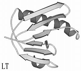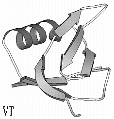

Verotoxin (VT) and Heat-labile Enterotoxin (LT) cause hemolytic uremia and traveller's diarrhea, respectively. From the 3D structures of both toxins it is clear that their B-subunits adopt the same fold, now known as the OB-fold. The RMS deviation of 47 CA is 1.0 Å while only three residues are identical in a structure-based sequence alignment (VT and LT B-subunit sequences are 69 and 101 residues long, respectively). Given the 3D structure of the VT B-subunit, can threading methods identify the fold of LT B?
Sequence alignment based on 3D structures
                                      b b b b b   b b b b b b
VT  - - - - - - - - - - - - - - - T P D C V T G K V E Y T K Y N D - - -
                                              | | | | | | | |
LT  A P Q T I T E L C S E Y R N T Q I Y T I N D K I L S Y T E S M A G K
            a a a a a           b b b b b b b b       b b b b b

          b b b b b       b b b b b                                   a
VT  D D T F T V K V - G D K E L F T N R - - - - - - - - - - - - - W N L
    | | | | | | | |       | | | | |                               | | |
LT  R E M V I I T F K S G E T F Q V E V P G S Q H I D S Q K K A I E R M
        b b b b b           b b b b                 a a a a a a a a a a

    a a a a a a a a a a     b b   b b b                       b b b b
VT  Q S L L L S A Q I T G M T V - T I K T N A C H N G G G F S E V I F R
    | | | | | | | | | | | | | |   | | | |           | | | | | | | | | |
LT  K D T L R I T Y L T E T K I D K L C V W N N K T P N S I A A I S M K N
    a a a a a a a a a         b b b b b b b           b b b b b b b b b
The OB-fold is:


10 best structures:
| PDB structure | raw score | Z-score | a.a.aligned | % identites | Fold (class) |
| 1bovAx 69 a.a. | 3062 | 10.40 | 69 | 100 | OB (all beta) |
| 1ltsDx 103 a.a. | 569 | 1.48 | 89 | 16 | OB (all beta) |
| 1mhcBx 99 a.a. | 543 | 1.38 | 81 | 20 | MHC recogn. dom. (alpha + beta) |
| 1ayaAx 101 a.a. | 470 | 1.12 | 79 | 21 | SH2-like(alpha + beta) |
| 1fkj_x 107 a.a. | 434 | 0.99 | 78 | 21 | FKBP-like (alpha + beta) |
| 2mcm_x 112 a.a. | 401 | 0.87 | 82 | 20 | Immunoglob.-like (all beta) |
| 1aac_x 105 a.a. | 386 | 0.82 | 84 | 26 | Cupredoxin-like(all beta) |
| 2fxb_x 81 a.a. | 381 | 0.80 | 75 | 13 | Ferredoxin-like (alpha + beta) |
| 1bdo_x 80 a.a. | 369 | 0.76 | 74 | 16 | ND |
| 1prtDx 110 a.a. | 366 | 0.75 | 84 | 17 | OB (all beta) |
Alignments:
1bovAx 3062 finds itself
     bb                     bbbbb      bbbbb    bb   bbaaaaa
MKKTLLIAASLSFFSASALATPDCVTGKVEYTKYNDDDTFTVKVGDKELFTNRWNLQSLL
                    ****************************************
--------------------TPDCVTGKVEYTKYNDDDTFTVKVGDKELFTNRWNLQSLL
                             bbbbb   bbbbbbbbbbbbbbb   aaaaa

aaa    bbbbbb         bbbbbbb
LSAQITGMTVTIKTNACHNGGGFSEVIFR
*****************************
LSAQITGMTVTIKTNACHNGGGFSEVIFR
aaaaaaabbbbbbb     bbbbb


1ltsDx 569 finds Heat-labile enterotoxin
     bb                           bb  bbb      bbbbb    bb
MKKTLLIAAS----LSFFS--ASALATPDCVTGKVE--YTKYNDDDTFTVKVGDKELF-T
  .*.    *     ..|.   . *  .:. .** *  ...|.:.|** *.*.:.:   .
APQTITELCSEYRNTQIYTINDKILSYTESMAGKREMVIITFKSGETFQVEVPGSQHIDS
   aaaaaaaa   bbbbbbb   bbbbbbb    bbbbbbbb bbbbb

  bbaaaaaaaa    bbbbbb             bbbbbbb
NRWNLQSLLLSAQITGMTVTIKTNAC----HNGGGFSEVIFR-
.. .::.:  . .** .* * . . *    ..:.:.... .|
QKKAIERMKDTLRITYLTETKIDKLCVWNNKTPNSIAAISMKN
aaaaaaaaaaaaaaaaaaabbbbb            bbbbbbb
--- ------------------------------------------------------------
--- TOPITS prediction-based threading
--- ------------------------------------------------------------
--- TOPITS ALIGNMENTS HEADER: ABBREVIATIONS
--- RANK         : rank in alignment list, sorted according to z-score
--- EALI         : alignment score
--- LALI         : length of alignment
--- IDEL         : number of residues inserted
--- NDEL         : number of insertions
--- ZALI         : alignment zcore;  note: hits with z>3 more reliable
--- PIDE         : percentage of pairwise sequence identity
--- LEN2         : length of aligned protein structure
--- ID2          : PDB identifier of aligned structure
--- NAME2        : name of aligned protein structure
---
--- TOPITS ALIGNMENTS HEADER: ACCURACY
---              : Tested on 80 proteins, TOPITS found the
---              : correct remote homologue in about 30% of
---              : the cases, detection accuracy was higher
---              : for higher z-scores (ZALI):
--- ZALI>0       : 1st hit correct in 33% of cases
--- ZALI>3       : 1st hit correct in 50% of cases
--- ZALI>3.5     : 1st hit correct in 60% of cases
---
--- TOPITS ALIGNMENTS HEADER: SUMMARY
 RANK   EALI LALI IDEL NDEL   ZALI PIDE LEN2  ID2 NAME2
    1  89.43   69    0    0   4.25  100  349 1bov_A VEROTOXIN-1 OB fold (all beta>
    2  88.48   69    0    0   4.18  100  349 1bov_A VEROTOXIN-1
    3  88.43   69    0    0   4.17  100  349 1bov_A VEROTOXIN-1
    4  88.28   69    0    0   4.16  100  349 1bov_A VEROTOXIN-1
    5  87.87   69    0    0   4.13  100  349 1bov_A VEROTOXIN-1
    6  57.28   88   14    5   1.82   19  476 2aaa D alpha-amylase beta dom (all beta)
    7  56.65   88    9    2   1.78   16  244 1rva_A restriction endonuclease (alpha/beta)
    8  55.65   86    4    2   1.70   16 1275 1tah_A alpha/beta hydrolase (alpha/beta)
    9  55.65   86    4    2   1.70   16 1275 1tah_A alpha/beta hydrolase (alpha/beta)
   10  55.48   87   15    3   1.69   15  405 1eft EF-Tu C-term Dom. (all beta)
   11  55.25   88   18    3   1.67   13  496 1smd L_ ND
   12  55.15   80    3    2   1.66   17 1275 1tah_A alpha/beta hydrolase (alpha/beta)
   13  54.92   83   14    4   1.65   17  131 1ifc Lipocalins (all beta)
   14  54.43   87    5    3   1.61   13  686 1cdg Immunoglobulin-like (all beta)
   15  54.10   87    6    4   1.58   22  648 2bbv_A Viral capsid proteins (all beta)
   16  53.93   86    4    2   1.57   16 1275 1tah_A alpha/beta hydrolase (alpha/beta)
   17  53.77   87   14    2   1.56   17  452 1gcb L_ID Immunoglobulin-like (all beta)
   18  53.37   81   13    4   1.53   15  377 1hsb_B Immunoglobulin-like (all beta)
   19  53.37   81   13    4   1.53   15  377 1hsb_A Immunoglobulin-like (all beta)
   20  53.28   86    6    4   1.52   22  648 2bbv_A Viral capsid proteins (all beta)
 The method used for this prediction was:  gonnet .
    NOTE: This method is merely a sequence-sequence comparison
          of your sequence to the sequences of the PDB folds.
Most similar fold:  1bova
 VEROTOXIN-1 (B-OLIGOMER, ALSO CALLED SHIGA-LIKE T

RANK Z-SCORE             FOLD LENGTHALI %ID
  1   49.98             1bova    69    100    2-21   beta ; OB-fold] 2-21-1-1-3-1-1-1
  2    4.23             1tiid    77     25    1997    new ; ESCHERICHIA COLI HEAT LABILE ENTEROTOXIN TYPE I]
  3    3.72        1dlha_3-81    72     21    4-12   (a+b) ; MHC antigen-recognition domain] 4-12-1-1-2-1-1-1 RC:,
  4    3.16              1ifl    52     15    1-71   alpha ; Oligomers of long helices] 1-71-3-1-1-4-1
  5    2.96              1drs    51     12    7-5    small ; Snake toxin-like] 7-5-1-2-1-1-1
  6    2.89              1erg    64     16    7-5    small ; Snake toxin-like] 7-5-1-3-1-1-1
  7    2.84     3hhrb_131-234    86     19    2-1    beta ; Immunoglobulin-like beta-sandwich] 2-1-2-1-5-1-1-2
  8    2.80              1mjc    63     24    2-21   beta ; OB-fold] 2-21-5-1-1-1-1
                         ---> Major cold shock protein from Escherichia coli (n=5, S=8)
  9    2.55              2cyr    72     26    7-27   small ; Cytochrome c3] 7-27-1-1-1-1-2
 10    2.42     1frta_179-269    64     23    2-1    beta ; Immunoglobulin-like beta-sandwich] 2-1-1-2-3-1-2-1

LEGEND:
COL. 1: RANK. The ranks are obtained by sorting the fold library,
              by Z-SCORES, in decreasing order. Only the 10
              structures that are most compatible to your sequence
              are shown.
COL. 2: Z-SCORE. The z-scores are  computed using
              the distribution of raw scores (not shown) of all folds.
COL. 3: FOLD. Protein Data Bank codes for the coordinates of the 3D
              structures.
COL. 4: LENGTHALI. The number of residues from your sequence that were
                   aligned to the fold.
COL. 5: % ID. Percentage of identical residues in the alignment.

RELIABILITY OF THIS PREDICTION:
           With this method the confidence threshold
           is a z-score of  4.4 +- 1.0.
![]() 5. Comparative Modeling: From Sequence to 3D model
5. Comparative Modeling: From Sequence to 3D model